Nikon Instruments announced the winners of the 51st annual Nikon Small World Photomicrography Competition, celebrating over five decades of excellence in microscopy and digital imaging:
1st Place
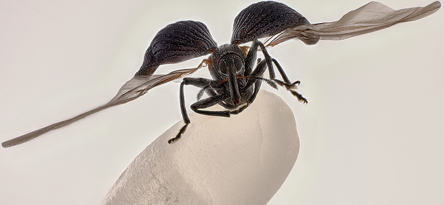
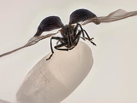
Zhang You, Kunming, Yunnan, China
Rice weevil (Sitophilus oryzae) on a grain of rice
Image Stacking 5X (Objective Lens Magnification)
2nd Place
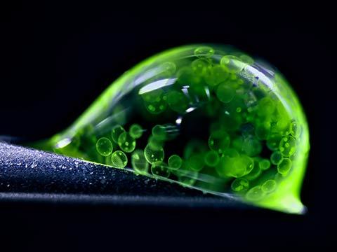
Dr. Jan Rosenboom, Rostock, Mecklenburg Vorpommern, Germany
Colonial algae (Volvox) spheres in a drop of water
Reflected Light 5X (Objective Lens Magnification)
3rd Place
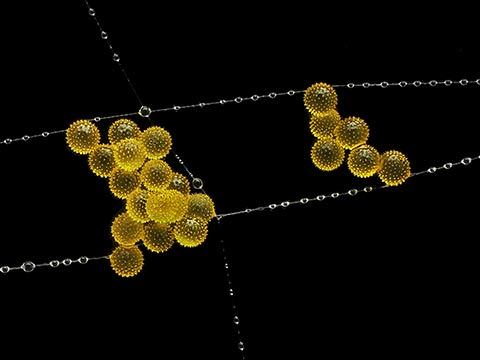
John-Oliver Dum, Medienbunker Produktion, Bendorf, Rheinland Pfalz, Germany
Pollen in a garden spider web
Image Stacking 20X (Objective Lens Magnification)
4th Place
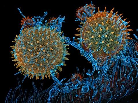
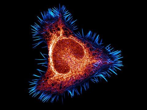
Dr. James Hayes, Vanderbilt University, Department of Cell and Developmental Biology, Nashville, Tennessee, USA
Heart muscle cells with chromosomes condensed following cell division
Confocal 100X (Objective Lens Magnification)
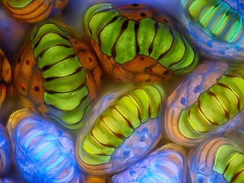
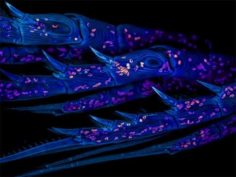
5th Place
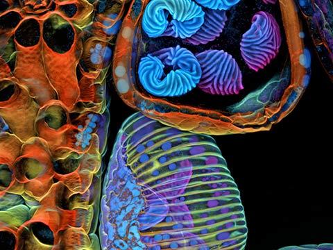
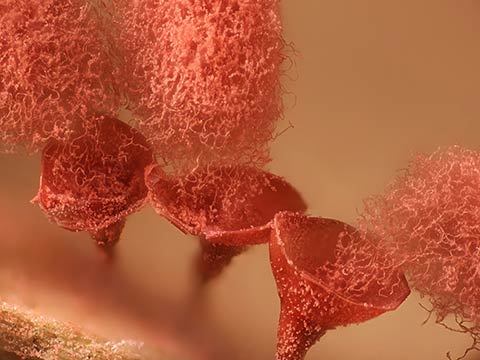
Dr. Igor Siwanowicz, Howard Hughes Medical Institute (HHMI), Janelia Research Campus, Ashburn, Virginia, USA
Spores (blue/purple structures) of a small tropical fern (Ceratopteris richardii)
Confocal 25X (Objective Lens Magnification)
6th Place
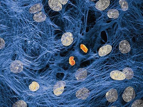
Dr. Francisco Lázaro-Diéguez, Albert Einstein College of Medicine, Bronx, New York, USA
Rat liver cells
Confocal 63X (Objective Lens Magnification)
7th Place
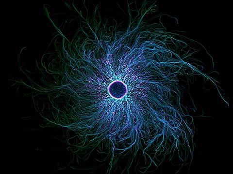
Stella Whittaker, National Institutes of Health, National Institute of Neurological Disorders and Stroke
Bethesda, Maryland, USA
iPSC-derived sensory neurons labelled to show tubulin and actin
Confocal, Fluorescence, Image Stacking, 10X (Objective Lens Magnification)
8th Place
Dr. Igor Siwanowicz, Howard Hughes Medical Institute (HHMI), Janelia Research Campus
Ashburn, Virginia, USA
Mallow pollen germinating on stigma while being parasitized by a filamentous fungus
Confocal, 40X (Objective Lens Magnification)
9th Place
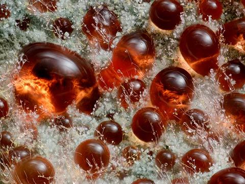
Wim van Egmond, Micropolitan Museum, Berkel en Rodenrijs, Zuid Holland, Netherlands
A fungus (Talaromyces purpureogenus) known for its red, diffused pigment
Image Stacking, 10X (Objective Lens Magnification)
10th Place
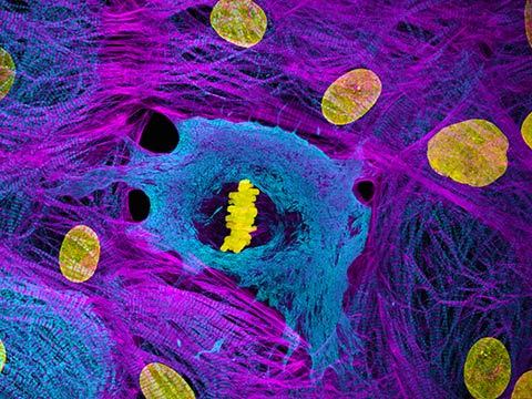
Dr. Dylan Burnette & Dr. James Hayes, Vanderbilt University School of Medicine
Department of Cell and Developmental Biology, Nashville, Tennessee, USA
Heart muscle cells (iPSC-derived) showing condensed chromosomes in metaphase
Structured Illumination Microscopy (SIM), 60X (Objective Lens Magnification)
11th Place
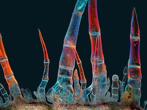
Marek Miś, Marek Miś Photography, Suwalki, Podlaskie, Poland
Sunflower trichomes (hair-like plant outgrowths)
Polarized Light, 10X (Objective Lens Magnification)
12th Place
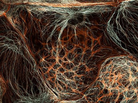
Halli Lindamood & Eric Vitriol, Augusta University, Department of Neuroscience and Regenerative Medicine
Augusta, Georgia, USA
The actin cytoskeleton (cyan) and endoplasmic reticulum (red) of a mouse brain cancer cell
Confocal, Deconvolution, 100X (Objective Lens Magnification)
13th Place
Henri Koskinen, Helsinki University, Helsinki, Uudenmaan lääni, Finland
Slime mold (Arcyria major) releasing spores
Image Stacking, Reflected Light, 10X (Objective Lens Magnification)
14th Place
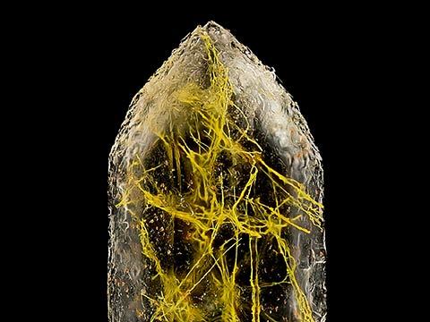
Manfred Heising, LWL Museum of Natural History Münster, Münster, Northrhine-Westphalia, Germany
Quartz with biotic goethite filaments
Image Stacking, 5X (Objective Lens Magnification)
15th Place
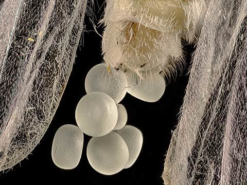
Zhang You, Kunming, Yunnan, China
Geometer moth (Geometridae) laying eggs
Image Stacking, 5X (Objective Lens Magnification)
16th Place
Rogelio Moreno, Panama, Panama
Spore sacs (sporangia) of a fern
Fluorescence, Image Stacking, 40X (Objective Lens Magnification)
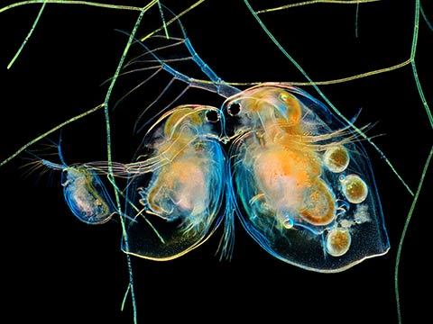
17th Place
Hong Guo, Chengdu, Si Chuan, China
Water fleas (Daphnia) and algae
Image Stacking, 5X (Objective Lens Magnification)
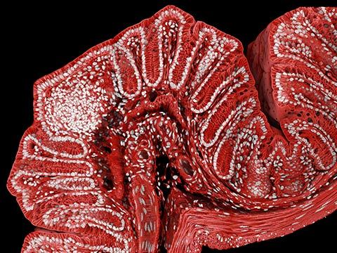
18th Place
Marius Mählen, Koen Oost, Prisca Liberali & Laurent Gelman, Friedrich Miescher Institute for Biomedical Research, Basel, Basel Stadt, Switzerland
Fluorescently marked mouse colon
Confocal, 20X (Objective Lens Magnification)
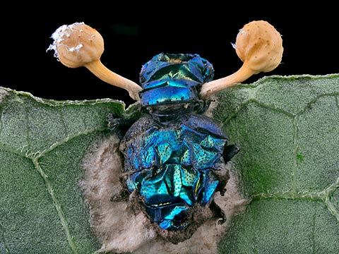
19th Place
Eduardo Agustin Carrasco, Cuenca, Azuay, Ecuador
Parasitic fungus (Cordycipitaceae) on a fly (Calliphoridae)
Image Stacking, 2X (Objective Lens Magnification)
20th Place
Zachary Sanchez, Vanderbilt University, Department of Cell and Developmental Biology
Nashville, Tennessee, USA
Marine copepod
Confocal, 60X (Objective Lens Magnification)
Nikon Small World Honors 51st Annual Photomicrography Competition
Nikon Instruments Inc. today announced the winners of the 51st annual Nikon Small World Photomicrography Competition, celebrating over five decades of excellence in microscopy and digital imaging. The first-place prize was awarded to China’s Zhang You for his striking image of a rice weevil mounted on a grain of rice. The image captures the insect with its wings fully extended, frozen in a moment that provides insight into the structure and behavior of a familiar yet often overlooked agricultural pest.
A member of the Entomological Society of China and the Entomological Society of Yunnan Province, You’s winning work is a product of the years he has spent focused on ecological and insect science photography, as well as teaching others about entomology. “It pays to dive deep into entomology: understanding insects’ behaviors and mastering lighting,” You said. “A standout work blends artistry with scientific rigor, capturing the very essence, energy, and spirit of these creatures.”
The choice of scale in the image emphasizes the insect’s actual size while contextualizing its ecological role as a pest known for attacking seeds of several crops. Using a medium-format camera paired with a 5x microscope objective, You captured over 100 images for focus stacking, carefully cleaning, lighting, and post-processing the specimen over the course of two weeks.
The subject itself was a rare and fortunate find. “I had observed rice weevils in grains before, but never one with its wings spread,” You explained. “This one was naturally preserved on a windowsill, perhaps in a final attempt to escape. Its tiny size makes manually preparing spread-wing specimens extremely difficult, so encountering it was both serendipitous and inspiring.” Insects, from pollinators to pests, play vital roles in ecosystems and economies alike, and You’s work encourages audiences to recognize the complexity hidden among these communities.
In addition to winning first place, You also earned 15th place in the 2025 competition with an image of a Geometer moth (Geomitridae) laying eggs, further demonstrating the range and depth of his skill.
“Zhang You’s work demonstrates the remarkable power of microscopy to reveal new perspectives on the world around us,” said Eric Flem, Senior Manager, Communications and CRM at Nikon Instruments. “What makes this year even more extraordinary is that it was his very first time entering the competition, and he not only captured first place, but also placed another image in the top 20. His achievement highlights the spirit of Nikon Small World: inspiring wonder, making scientific understanding accessible to all, and celebrating the artistry of the microscopic realm.”