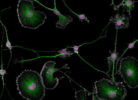
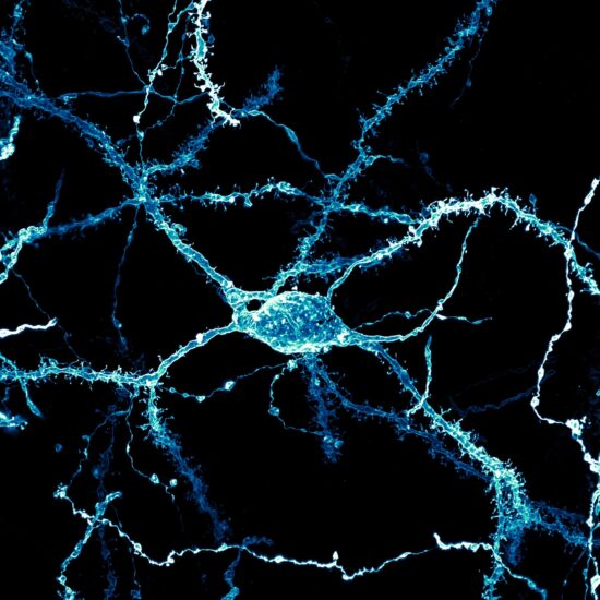
First Place, 2024 Nikon Small World Photomicrography Competition Differentiated mouse brain tumor cells (actin, microtubules, and nuclei)
Nikon announced the winners of the 2024 Small World Photo Awards.
Additional information:

Nikon Instruments Inc. today announced the winners of the 50th annual Nikon Small World Photomicrography Competition, celebrating five decades of excellence in microscopy and digital imaging. This year’s first place prize was awarded to Dr. Bruno Cisterna, with assistance from Dr. Eric Vitriol of Augusta University, for his groundbreaking image of differentiated mouse brain tumor cells, highlighting the actin cytoskeleton, microtubules, and nuclei. This image reveals how disruptions in the cell’s cytoskeleton – the structural framework and “highways” known as microtubules – can lead to diseases like Alzheimer’s and ALS.
Dr. Cisterna’s research revealed that profilin 1 (PFN1), a protein crucial for building the cell’s structure, plays a key role in maintaining the microtubule highways essential for cellular transport. When PFN1 or related processes are disrupted, these highways can malfunction, leading to cellular damage similar to what is observed in neurodegenerative diseases.
“One of the main problems with neurodegenerative diseases is that we don’t fully understand what causes them,” said Dr. Cisterna. “To develop effective treatments, we need to figure out the basics first. Our research is crucial for uncovering this knowledge and ultimately finding a cure. Differentiated cells could be used to study how mutations or toxic proteins that cause Alzheimer’s or ALS alter neuronal morphology, as well as to screen potential drugs or gene therapies aimed at protecting neurons or restoring their function.”
His patience and determination were crucial in capturing his image. “I spent about three months perfecting the staining process to ensure clear visibility of the cells. After allowing five days for the cells to differentiate, I had to find the right field of view where the differentiated and non-differentiated cells interacted. This took about three hours of precise observation under the microscope to capture the right moment, involving many attempts and countless hours of work to get it just right.”
The hard work behind this discovery underscores its significance, bringing researchers closer to answers that could potentially transform millions of lives. “After three years of research, we finally published our findings four months ago in the Journal of Cell Biology, and there’s still more work to be done,” said Dr. Cisterna. “I’m deeply passionate about scientific imaging; I’ve been following the Nikon Small World contest for about 15 years. It’s an incredible contest that highlights the beauty of photomicrography but also inspires continued exploration and innovation in the field.”
Eric Flem, Senior Manager, CRM and Communications at Nikon Instruments, shares a similar perspective on the competition. “At 50 years, Nikon Small World is more than just an imaging competition – it’s become a gallery that pays tribute to the extraordinary individuals who make it possible. They are the driving force behind this event, masterfully blending science and art to reveal the wonders of the microscopic world and what we can learn from it to the public.” He went on to add, “Sometimes, we overlook the tiny details of the world around us. Nikon Small World serves as a reminder to pause, appreciate the power and beauty of the little things, and to cultivate a deeper curiosity to explore and question.”
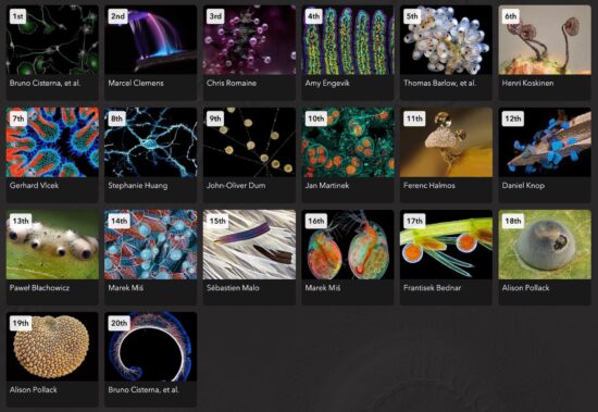
NIKON SMALL WORLD WINNERS
1st Place
Dr. Bruno Cisterna & Dr. Eric Vitriol
Medical College of Georgia at Augusta University
Department of Neuroscience & Regenerative Medicine
Augusta, Georgia, USA
Differentiated mouse brain tumor cells (actin, microtubules, and nuclei)
Super-Resolution
100X (Objective Lens Magnification)
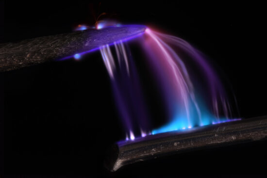
2nd Place
Dr. Marcel Clemens
Verona, Veneto, Italy
Electrical arc between a pin and a wire
Image stacking for the pin and wire combined with long exposure for the electrical arcs
10X (Objective Lens Magnification)
3rd Place
Chris Romaine
Kandid Kush
Port Townsend, Washington, USA
Leaf of a cannabis plant. The bulbous glands are trichomes. The bubbles inside are cannabinoid vesicles.
Image Stacking
20X (Objective Lens Magnification)
4th Place
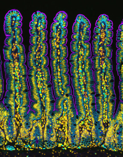
Dr. Amy Engevik
Medical University of South Carolina
Department of Regenerative Medicine & Cell Biology
Charleston, South Carolina, USA
Section of a small intestine of a mouse
Fluorescence
10X (Objective Lens Magnification)
5th Place
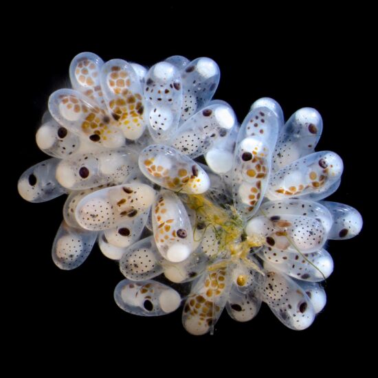
Thomas Barlow & Connor Gibbons
Columbia University
Department of Neurobiology and Behavior
New York, New York, USA
Cluster of octopus (Octopus hummelincki) eggs
Darkfield, Stereomicroscopy, Focus Stacking
3X (Objective Lens Magnification)
6th Place
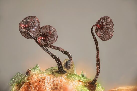
Henri Koskinen
Helsinki University
Helsinki, Uudenmaan lääni, Finland
Slime mold (Cribraria cancellata)
Image Stacking, Polarized Light, Reflected Light
10X (Objective Lens Magnification)
7th Place
Gerhard Vlcek
Maria Enzersdorf, Austria
Cross section of European beach grass (Ammophila arenaria) leaf
Brightfield, Image Stacking
10X (Objective Lens Magnification)
8th Place
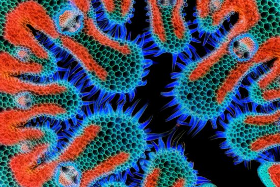
Stephanie Huang
Victoria University of Wellington
School of Biological Sciences; School of Psychology
Wellington, New Zealand
A neuron densely covered in dendritic spines from the striatum of an adult rat brain
Confocal, Deconvolution, Image Stacking
60X (Objective Lens Magnification)
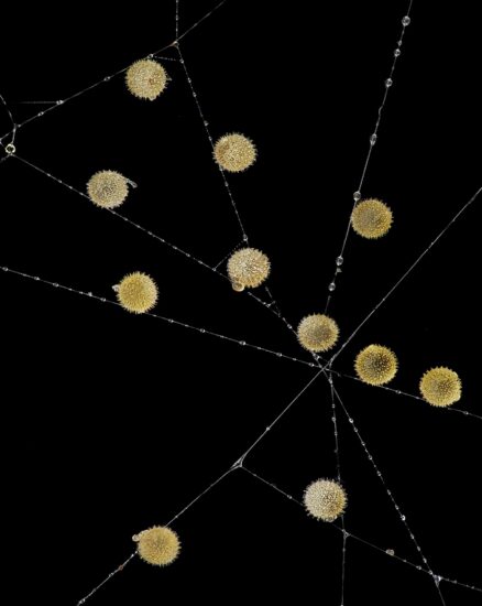
9th Place
John-Oliver Dum
Medienbunker Produktion
Bendorf, Rheinland Pfalz, Germany
Pollen in a garden spider (Araneus) web
Image Stacking
20X (Objective Lens Magnification)

10th Place
Jan Martinek
Charles University
Department of Experimental Plant Biology
Prague, Czech Republic
Spores of black truffle (Tuber melanosporum)
Confocal
63X (Objective Lens Magnification)
Congratulations to all the winners of our milestone 50th anniversary #NikonSmallWorld competition! We’re so grateful for every single person who submitted and made this competition into what it is today. The complete 2024 winning gallery is now live: https://t.co/I8Pcou7949 🔬 pic.twitter.com/YDWeSA99GD
— Nikon Small World (@NikonSmallWorld) October 17, 2024
![]()
![]()